<script type=”application/ld+json”>
{
“@context”: “https://schema.org”,
“@graph”: [
{
“@type”: “Service”,
“name”: “Retinal Detachment Surgery”,
“serviceType”: “Retinal Surgery, Vitrectomy, Scleral Buckling”,
“provider”: {
“@type”: “Organization”,
“name”: “Centre for Sight”,
“url”: “https://www.centreforsight.com”,
“logo”: “https://www.centreforsight.com/wp-content/uploads/2023/03/Centre-for-Sight-Logo.svg”
},
“areaServed”: {
“@type”: “Place”,
“name”: “United Kingdom”
},
“description”: “Centre for Sight provides expert surgical treatment for retinal detachment, using advanced techniques such as vitrectomy and scleral buckling to preserve and restore vision. Our experienced team of retinal specialists ensures world-class outcomes in a patient-centred environment.”,
“image”: “https://www.centreforsight.com/wp-content/smush-webp/2024/05/laser-eye-surgery-young-man-smiling-1481×1536.jpg.webp”
},
{
“@type”: “Product”,
“name”: “Retinal Detachment Surgery”,
“description”: “Centre for Sight offers retinal detachment surgery performed by expert retinal surgeons using state-of-the-art technology to restore retinal attachment and prevent vision loss.”,
“image”: “https://www.centreforsight.com/wp-content/smush-webp/2024/05/laser-eye-surgery-young-man-smiling-1481×1536.jpg.webp”,
“brand”: {
“@type”: “Brand”,
“name”: “Centre for Sight”
},
“review”: {
“@type”: “Review”,
“reviewRating”: {
“@type”: “Rating”,
“ratingValue”: “5”,
“bestRating”: “5”
},
“author”: {
“@type”: “Person”,
“name”: “mbb”
},
“reviewBody”: “Mr Saj Khan recently removed the cataract in my left eye and replaced it with a trifocal lens. I am delighted with the result and very happy with the care I received from the team at Centre for Sight. …More”
},
“aggregateRating”: {
“@type”: “AggregateRating”,
“ratingValue”: “4.1”,
“reviewCount”: “44”,
“bestRating”: “5”
}
}
]
}
</script>
Retinal Detachment Surgery
Retinal detachment is a serious condition of the eye in which the retina stops receiving oxygen. The symptoms of a retinal detachment can be frightening. Objects might appear to float across your eye, or a grey veil may move across your field of vision.
If not treated quickly, a retinal detachment can cause you to lose your vision permanently. Retinal detachment repair is a surgical procedure that is used to restore circulation to the retina and preserve vision.
Retinal Detachment Surgery Overview
The retina is the lining of the back of the eye, which allows the eye to see. If a hole appears in the retina it will detach rather like wallpaper peeling off a wall. The retina cannot work when it is detached. The only way to repair the retina is by an operation to find and seal any holes. This may take the form of a simple laser procedure or freezing (cryotherapy) if caught early enough. If the retina is detached, a procedure called a vitrectomy may be required in addition to laser and/or cryotherapy.
Certain symptoms may indicate retinal detachment which , these include the onset of new floaters, loss of vision a shadow or curtain of visual loss, visual distortion, flashes and floaters, blurred vision and / or patchy vision.
Almost all patients with retinal detachments must have surgery to place the retina back in its proper position. Otherwise, the retina will lose the ability to function, possibly permanently, and blindness can result. The method for fixing retinal detachment depends on the characteristics of the detachment. In each of the following methods, your ophthalmologist will locate the retinal tears and use laser surgery or cryotherapy to seal the tear.
Read More
Information about Retinal Detachment
Symptoms
Before the actual retinal detachment occurs there is often a tear in the retina. This may present with the following symptoms:
Light flashes, which occurs mainly with jerky eye movements, or in the dark. (Light flashes and dots which occur as a result of fluctuations in blood pressure, for example when standing up or re-erecting after bending, are harmless!)
Semi-transparent streaks that “migrate” with eye movements. These are usually so-called “Mouches volantes” which are annoying but not dangerous, however should always be checked by your consultant ophthalmologist
A “rain of soot”, the perception of a large amount of small black dots or floating particles sinking downwards within your eye
These symptoms can be indicative of the retina being torn. Such a tear in the retina can be “repaired” by means of a laser, similar to a weld – a retinal scar remains and at this position a very small visual field defect that typically stays unnoticed.
When the retina detaches itself in the area of the macula, central vision is compromised: with loss of clarity and detail with only blurred and distorted vision remaining.
Retinal detachment does not cause any pain. Untreated retinal detachment can lead to blindness of the eye affected.
The vitreous body shrinks with increasing age and may lift off the retina. In areas where it is conjoined with the retina, a tear might then develop
Blunt force trauma e.g. an object or a child’s finger bouncing against the eye
Result of a cataract-operation
Inflammatory processes in the choroid membrane, e.g. diabetic retinopathy in cases of diabetes mellitus
Treatment for Retinal Detachment
There are several types of surgery to repair a detached retina. A simple tear in the retina can be treated with freezing, called cryotherapy, or a laser procedure. Different types of retinal detachment require different kinds of surgery and different levels of anaesthesia. The type of procedure your Consultant Ophthalmic Surgeon performs will depend on the severity of retinal detachment.
The Consultation
Following a detailed assessment, your Consultant Ophthalmic Surgeon will discuss the best course of treatment for you. The consultant will be the same person who carries out your procedure and your checks at follow-up appointments.
The consultation process is very detailed and can take up to 2 hours. Tests and investigations are performed by nurses, optometrists and technicians and your pupils will be dilated with eye drops. You will be provided further information about the procedure and shown video animations.
You will then be seen by a very experienced fellowship-trained consultant who will evaluate your eyes and come to a final decision. The consultant will inform you about your suitability and what options there are for lenses as well as explain the process further and expectation of outcomes following surgery. You will be provided with an informed consent.
The Procedure
Depending on the position and extent of the detachment, there are different operative options, either externally or internally to the eye.
The aim is to reattach the retina.
The retina can be repaired in one of two ways:
- The hole can be sealed by sewing a small piece of plastic onto the outside of the eye, creating a dent in the eye ball, which will close the hole. You may be able to feel the plastic on the eye after the operation.
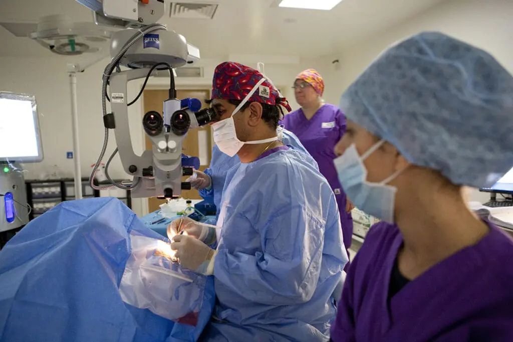
- Alternatively, it is possible to go inside the eye and, by removing the jelly in the eye (known as the vitreous), a gas bubble may be inserted to support the retina. This procedure is known as a Vitrectomy and is performed by making three small incisions in the eye. The operation takes approximately one hour to perform. The gas bubble will float inside the eye and close the hole. Laser or freezing treatment is used to seal the hole.
The surgeon will decide which type of surgery is most appropriate for each individual patient. The eye does not need the vitreous jelly – it will fill the space with a watery fluid.
Aftercare and recovery
Your consultant ophthalmologist will give you eye drops to reduce any inflammation and to prevent infection. Please don’t rub your eye as this may increase infection and lead to complications.If you experience discomfort, we suggest that you take a pain reliever, such as paracetamol – take care not to exceed the dose stated on the packaging. It is normal to feel itching, and have sticky eyelids and mild discomfort in the operated eye for five to ten days following retinal detachment surgery. If you have severe pain that persists, you must call your consultant ophthalmologist.
It is also common for some fluid to leak from around your eye. Occasionally, the area surrounding your eyes can become slightly bruised – this is especially common after a scleral buckle procedure. Any discomfort should ease after one to two days.
In most cases, your eye will take about two to six weeks to heal. We will make an appointment for you to see your doctor again, usually within seven to 14 days of your operation. Try to rest while your eye is healing.
Complications / Risks
A retinal detachment can occur at any age, but it is more common in people over age 40. Every patient is different and some detached retinas are more complicated to treat than others. Some patients might need more than one operation.There is an 85-90% success rate with one operation of your retina going flat and staying flat. There is a 5-10% risk that you will need further surgery due to new breaks forming in your retina or the development of scar tissue.
Possible complications could be –
- Bruising of the eye or eyelids
High pressure inside the eye
Inflammation inside the eye
Cataract
Double vision
Allergy to the medication used
Infection in the eye (endophthalmitis) – this is very rare, but can lead to serious loss of sight
When can I drive again and do I need to inform the DVLA of my surgery?
When you can drive again will depend on the vision in your un-operated eye. We will assess this when you attend the postoperative clinic. You will be advised whether you will need to contact the DVLA at this appointment.
What should I do if I get pain in my eye?
It is normal to feel some discomfort after your surgery. This should be relieved by taking regular pain-killers, such as paracetamol. If you experience severe pain in your eye, please contact us on 0808 303 8601.
How long does it take for the redness in my eye to go?
Generally redness takes a few weeks to settle. The eye is red as a result of the surgery and this is entirely normal during the post-operative period.


